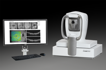
OCT-Angiography
|
Optical Coherence Tomography Angiography (OCTA) is a new non-invasive way to visualize the blood vessels in the back of your eye down to a capillary level. This enables the earliest detection of eye diseases (such as glaucoma and macular diseases) which affect the microscopic blood vessels not visible with older technology instruments. OCTA also reduces the need for the more invasive and higher risks of Fluorescein Angiography. Our OCTA allows the correct diagnosis of glaucoma, macular degeneration, diabetic retinopathy and other macula diseases.
|
Glaucoma Detection
Glaucoma is an eye disease of the optic nerve and retinal nerve tissue. The cause is believed to be from increased eye pressure, decreased retinal blood flow (oxygen supply) or a combination of both. Standard Optical Coherence Tomography (OCT) can measure thinning and therefore damage to the optic nerve and retinal nerve tissue. OCTA also allows imaging of reduced blood vessel density which indicates the presence of early glaucoma disease.
Macular Degeneration
Macular degeneration is also a disease which is best diagnosed with OCTA. Macular degeneration causes a loss of central vision due to damage of the macula. There are two types of macular degeneration, dry and wet. Wet macular degeneration occurs when abnormal blood vessels grow under the retina. These abnormal blood vessels are much weaker than normal ones and can rupture causing bleeding and swelling of the retina or macula. This results in significant vision loss and distortion. The good news is now with OCTA the abnormal blood vessels can be found earlier than ever before. Wet macular degeneration also can be treated so if diagnosed early, and treated early, often vision can improve.
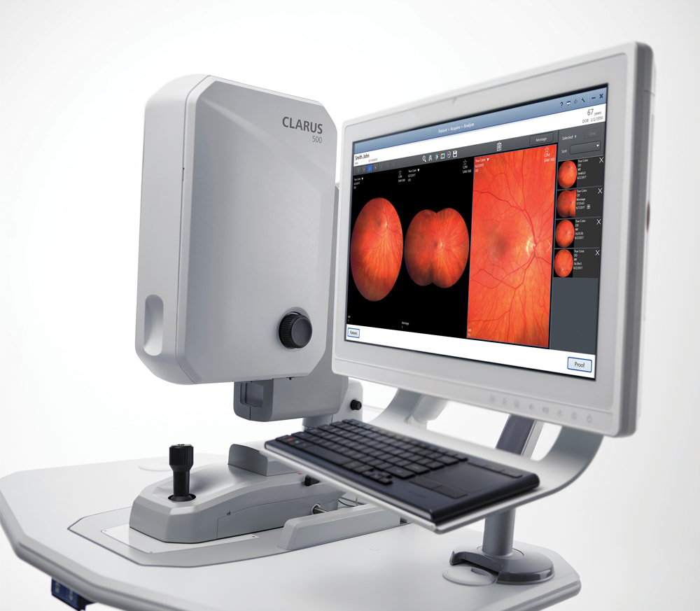
Clarus Ultrawide Field
|
The Zeiss Clarus allows our clinic to capture extremely clear and full field views of the internal eye. The instrument is an ultra-widefield camera that captures high resolution images down to 7 microns.
What this means for our patients is that we are able to document and observe all manner of ocular disease and track subtle changes in pathology over time. In addition, it also captures detailed fundus auto-florescence images and external structures as well. Often these capabilities are enjoyed without having to dilate our patients allowing comprehensive eye care with little to no inconvenience. . |
|